
Understanding Multiple Sclerosis: Causes and Symptoms
Multiple Sclerosis (MS) Explained in Plain English Imagine your nerves are like electrical wires with rubber coating (called myelin).In MS, […]
Multiple sclerosis (MS) diagnosis has changed a lot in the past few decades — and the timeline is pretty clear.
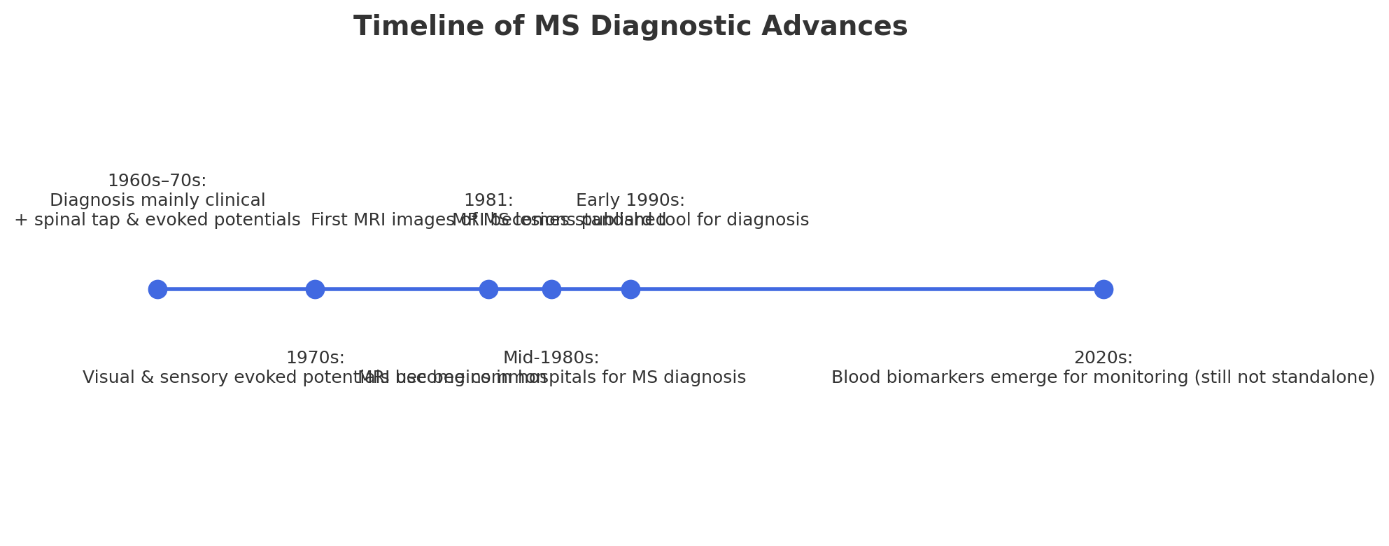
This method could take years to confirm MS because doctors had to wait for multiple flare-ups affecting different areas.
✅ Bottom line:
Multiple Sclerosis (MS) Explained in Plain English Imagine your nerves are like electrical wires with rubber coating (called myelin).In MS, […]
A well-balanced, anti-inflammatory diet can be beneficial for managing Multiple Sclerosis (MS). It supports immune function. It helps reduce flare-ups
Living with Multiple Sclerosis (MS) while on Ocrevus (ocrelizumab) requires a comprehensive approach. This includes medication management. It also involves
1. Seated Balance Exercises (Start Here) (Build core/leg strength before standing drills.) 2. Standing Balance Drills (With Walker Support) (Always
1. If You Have Relapsing-Remitting MS (RRMS): Red Flags (RRMS): 2. If You Have Primary Progressive MS (PPMS): Red Flags
Of course! If you’re looking for general help with multiple sclerosis (MS), here’s a breakdown of key areas to consider.
Typical Ocrevus Infusion Schedule & When Benefits May Appear: Factors That Influence Response: When to Expect Noticeable Changes? Important Notes: